44 fluorescent labels and light microscopy
How to Prepare Your Specimen for Immunofluorescence Microscopy Jan 10, 2022 · Immunofluorescence (IF) is a powerful method for visualizing intracellular processes, conditions and structures. IF preparations can be analyzed by various microscopy techniques (e.g. CLSM, Epifluorescence, TIRF, GSDIM), depending on the application or the researcher’s interest.Meanwhile, IF has become indispensable for a large number of research … ZEISS LSM 980 with Airyscan 2 NIR fluorescent labels are less phototoxic for living samples due to the longer wavelength. This allows you to investigate living samples for longer periods of time while limiting the influence of light. Additionally, light of longer wavelength ranges is less scattered by the sample tissue, increasing penetration depth.
Two-photon excitation microscopy - Wikipedia Two-photon excitation microscopy (TPEF or 2PEF) is a fluorescence imaging technique that allows imaging of living tissue up to about one millimeter in thickness, with 0.64 μm lateral and 3.35 μm axial spatial resolution. Unlike traditional fluorescence microscopy, in which the excitation wavelength is shorter than the emission wavelength, two-photon excitation requires …
Fluorescent labels and light microscopy
Fluorescence - Wikipedia Fluorescence is the emission of light by a substance that has absorbed light or other electromagnetic radiation.It is a form of luminescence.In most cases, the emitted light has a longer wavelength, and therefore a lower photon energy, than the absorbed radiation.A perceptible example of fluorescence occurs when the absorbed radiation is in the ultraviolet region of the … Super-resolution microscopy - Wikipedia Super-resolution microscopy is a series of techniques in optical microscopy that allow such images to have resolutions higher than those imposed by the diffraction limit, which is due to the diffraction of light. Super-resolution imaging techniques rely on the near-field (photon-tunneling microscopy as well as those that utilize the Pendry Superlens and near field scanning optical … Förster resonance energy transfer - Wikipedia Förster resonance energy transfer (FRET), resonance energy transfer (RET) or electronic energy transfer (EET) is a mechanism describing energy transfer between two light-sensitive molecules (chromophores). A donor chromophore, initially in its electronic excited state, may transfer energy to an acceptor chromophore through nonradiative dipole–dipole coupling.
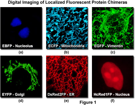
Fluorescent labels and light microscopy. Expansion microscopy: principles and uses in biological research Dec 20, 2018 · After gelation, digestion, and expansion, the fluorescence of the antibody labels or fluorescent proteins is largely retained for many genetically encoded fluorophore and small-molecule dyes (Fig ... (PDF) Introduction to Microscopy - ResearchGate Nov 08, 2017 · 1. Microscopy with light and electrons 2. Electron/specimen interactions: processes and detectors 3. The electron microscope family 4. Specimen preparation for electron microscopy 5. cellSens | Imaging Software | Olympus LS - Life Science Olympus cellSens software provides an intuitive imaging experience and seamless workflow for evolving research needs. Find out more. Introduction to Fluorescent Proteins | Nikon’s MicroscopyU The brightness and fluorescence emission spectrum of enhanced yellow fluorescent protein combine to make this probe an excellent candidate for multicolor imaging experiments in fluorescence microscopy. Enhanced yellow fluorescent protein is also useful for energy transfer experiments when paired with enhanced cyan fluorescent protein (ECFP) or ...
Förster resonance energy transfer - Wikipedia Förster resonance energy transfer (FRET), resonance energy transfer (RET) or electronic energy transfer (EET) is a mechanism describing energy transfer between two light-sensitive molecules (chromophores). A donor chromophore, initially in its electronic excited state, may transfer energy to an acceptor chromophore through nonradiative dipole–dipole coupling. Super-resolution microscopy - Wikipedia Super-resolution microscopy is a series of techniques in optical microscopy that allow such images to have resolutions higher than those imposed by the diffraction limit, which is due to the diffraction of light. Super-resolution imaging techniques rely on the near-field (photon-tunneling microscopy as well as those that utilize the Pendry Superlens and near field scanning optical … Fluorescence - Wikipedia Fluorescence is the emission of light by a substance that has absorbed light or other electromagnetic radiation.It is a form of luminescence.In most cases, the emitted light has a longer wavelength, and therefore a lower photon energy, than the absorbed radiation.A perceptible example of fluorescence occurs when the absorbed radiation is in the ultraviolet region of the …
The Biological bulletin. Biology; Zoology; Biology; Marine ...
Labeling proteins inside living cells using external ...
Frontiers | Label-Free Non-linear Multimodal Optical ...

Immunofluorescence Microscopy. Immunofluorescence Microscopy ...

Chemogenetic Tags with Probe Exchange for Live-Cell ...

VIPER is a genetically encoded peptide tag for fluorescence ...
light-microscopy-fluorescent-labels-neuroscience-shutterstock ...

In Silico Labeling: Predicting Fluorescent Labels in ...
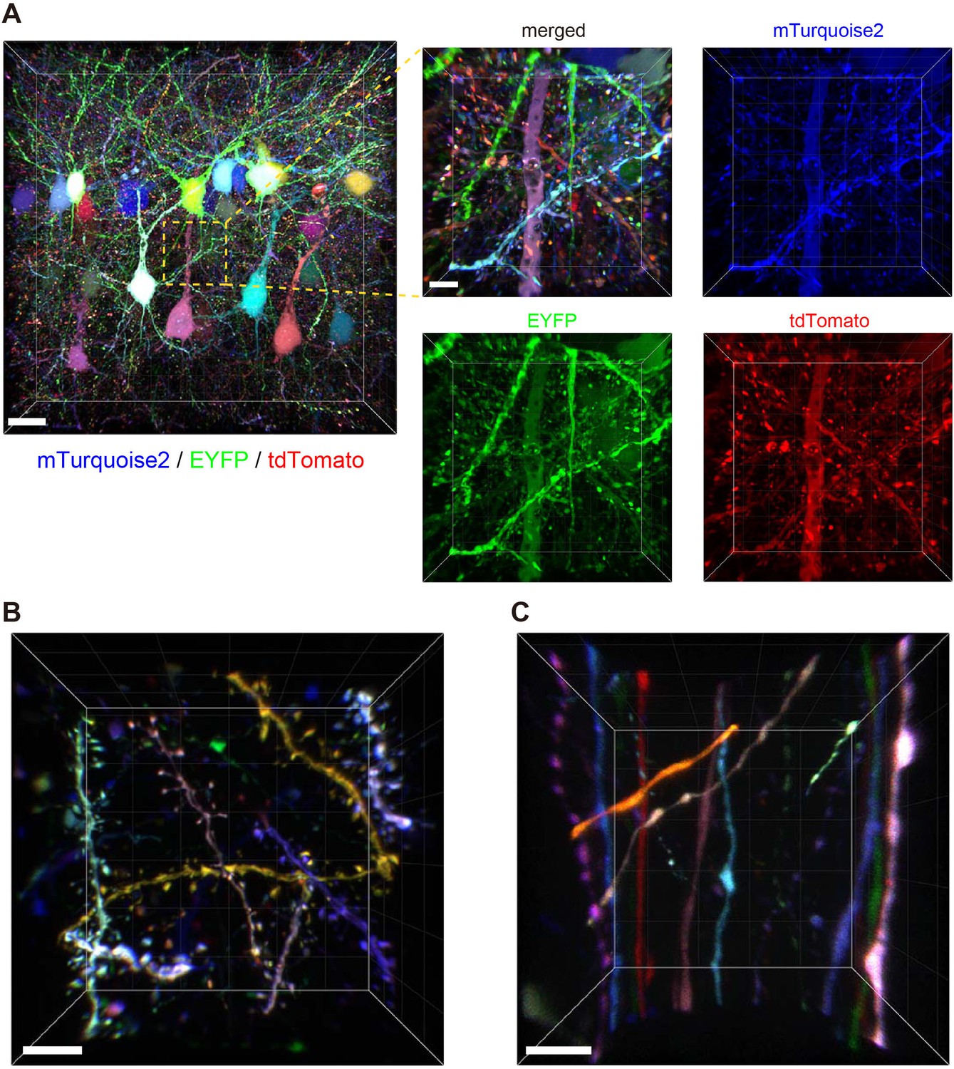
Bright multicolor labeling of neuronal circuits with ...

Microscopy: Intro to microscopes & how they work (article ...
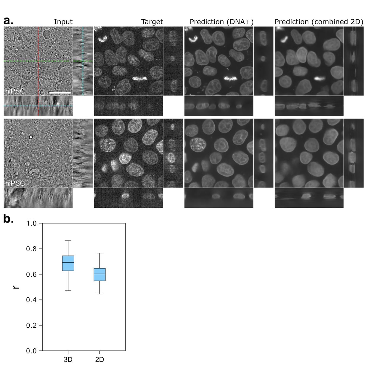
Label-free prediction of three-dimensional fluorescence ...

From Fluorophores to Filters: A guide to Fluorescence Microscopy
In Silico Labeling: Predicting Fluorescent Labels in ...
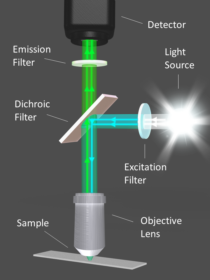
Fluorescence Filters for Microscopy and Imaging - Alluxa
Fluorescence Microscopy vs. Light Microscopy

ZEISS Axioscope 5 Smart Laboratory Microscope

Genetically encoded fluorescent tags | Molecular Biology of ...
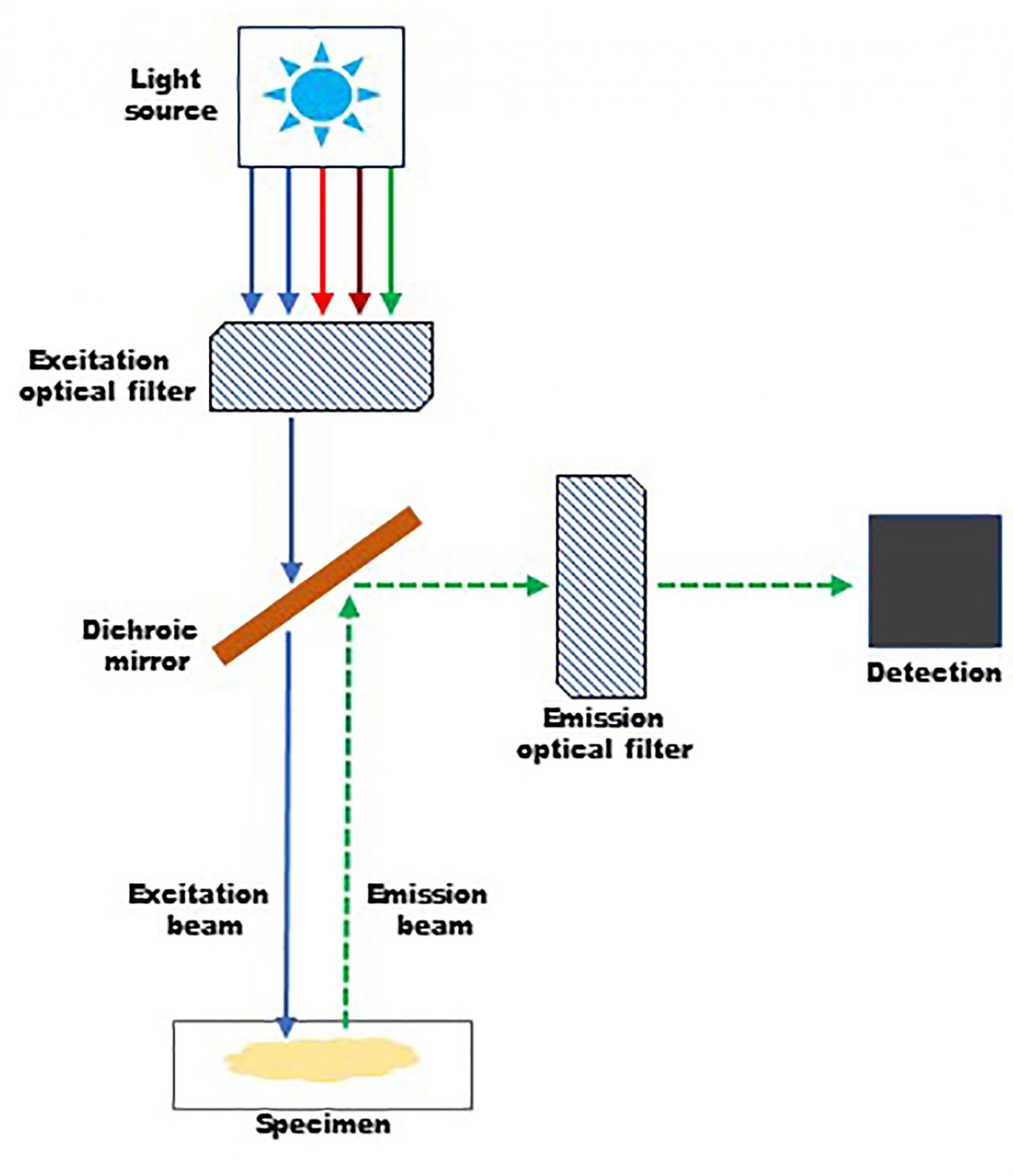
An Overview of Fluorescence Microscopy - Biotium
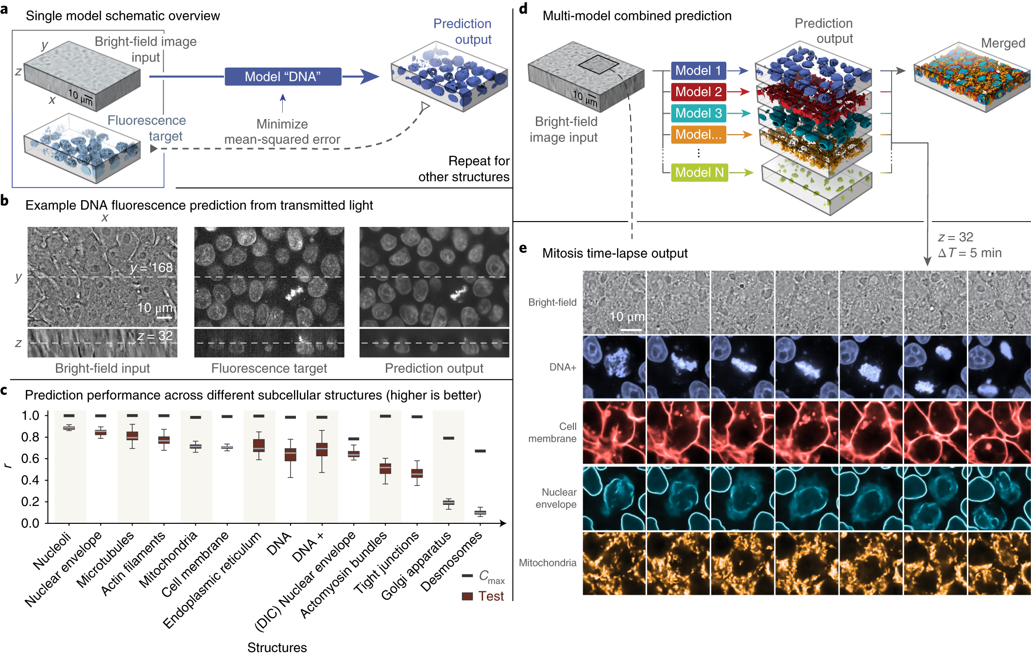
Label-free prediction of three-dimensional fluorescence ...
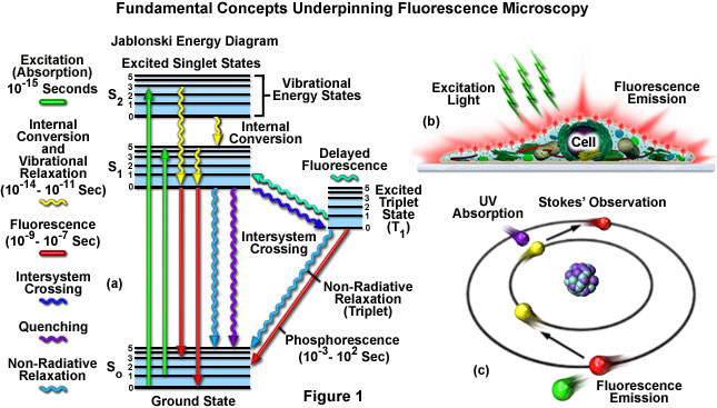
Definition > Fluorescence microscope

Google AI Blog: Seeing More with In Silico Labeling of ...
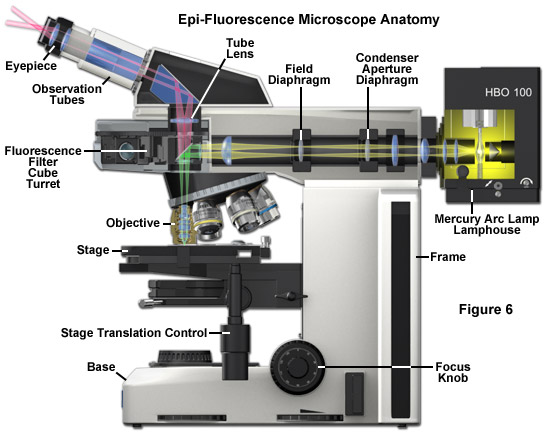
ZEISS Microscopy Online Campus | Microscopy Basics ...

VIPER is a genetically encoded peptide tag for fluorescence ...
Super-resolution microscopy - Wikiwand

How do you choose the right fluorophore? - Enzo Life Sciences
Making quantitative time-resolved fluorescence microscopy ...
Molecular Expressions Microscopy Primer: Specialized ...

The Microscope
Fluorescence microscope - Wikipedia

Fluorophores and Optical Filters for Fluorescence Microscopy

Single-cell cytometry via multiplexed fluorescence prediction ...

Fluorescent Labelling, FRET & Light Sheet Microscopy – Ben Sutcliffe
Fluorescence Microscopy - Explanation and Labelled Images ...

Fluorescent protein tags | Proteintech Group

Light microscopes — Science Learning Hub
Correlative fluorescence microscopy, transmission electron ...
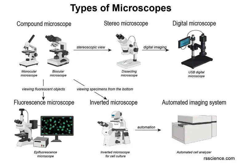
Different types of Microscopes – light microscope, electron ...
ZEISS Microscopy Online Campus | Microscopy Basics ...
Covalent labeling of nucleic acids - Chemical Society Reviews ...
Light Microscopy Techniques Used for Virology- Oxford Instruments
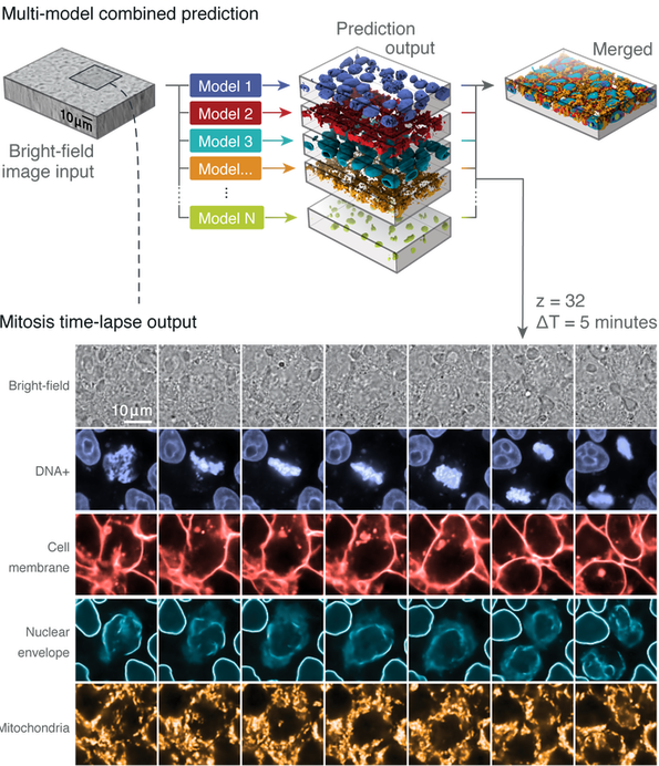
Label-free Determination - ALLEN CELL EXPLORER
Advanced Light Microscopy Methods - 2008 - Wiley Analytical ...

Fluorescence Images without Fluorescent Markers | AICS Modeling
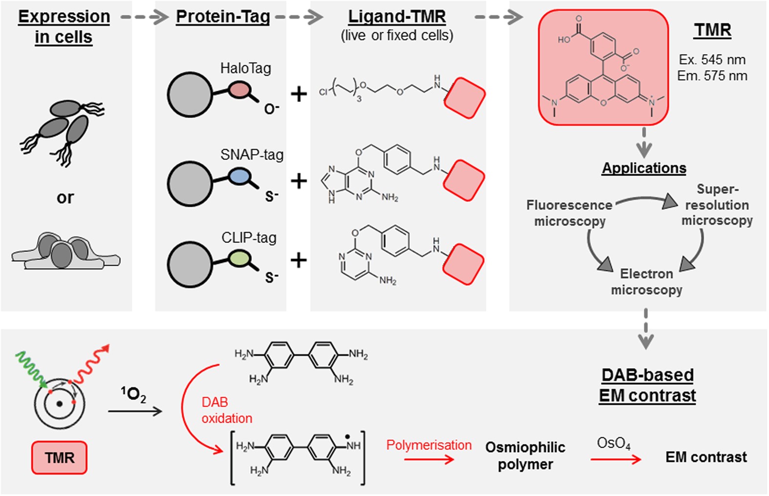
Self-labelling enzymes as universal tags for fluorescence ...
Post a Comment for "44 fluorescent labels and light microscopy"