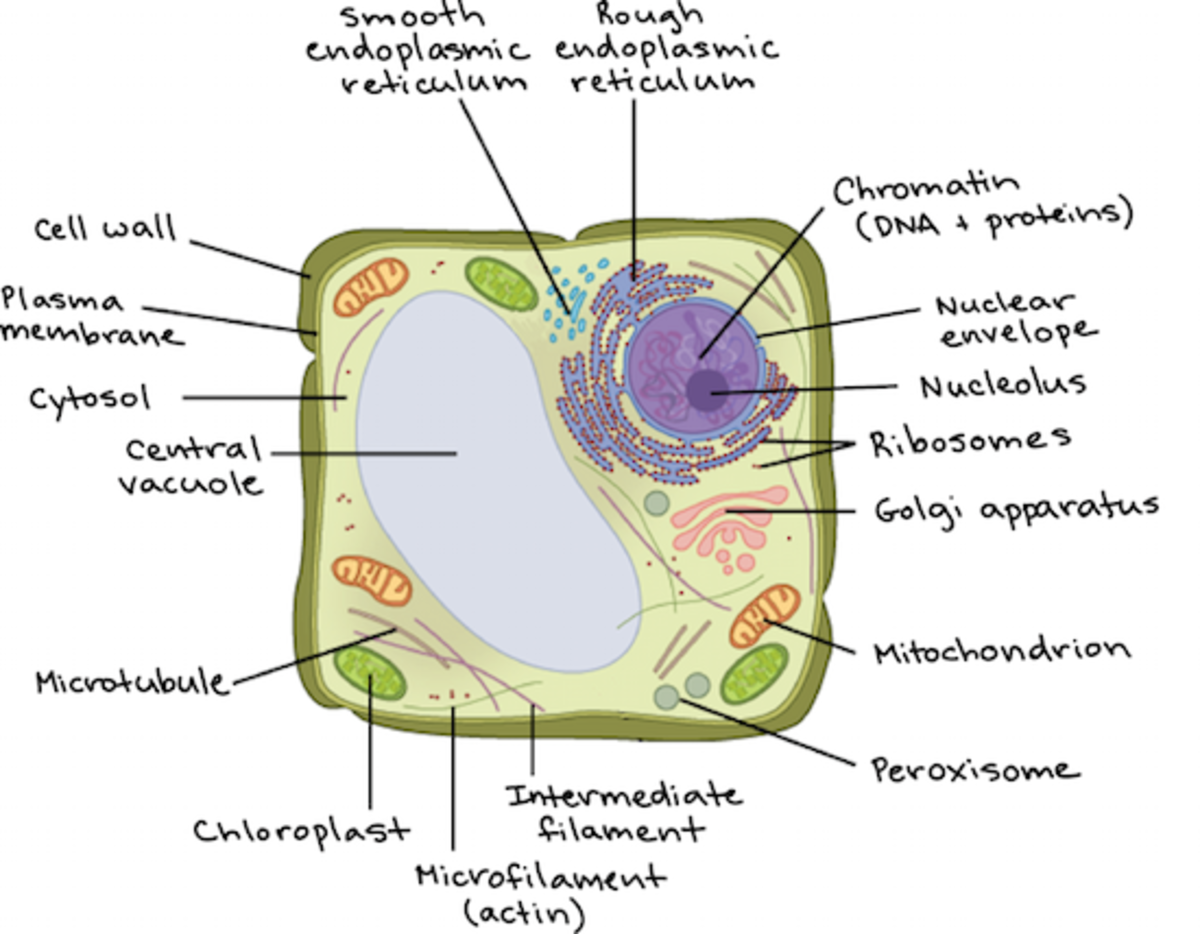
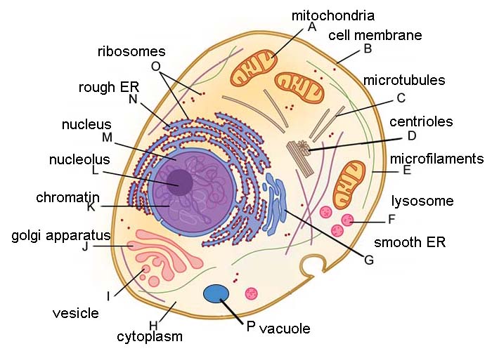
42 diagram of a cell with labels
Cell Diagrams - The Biology Corner Open Google Draw and import the diagram. Then use "insert" to create text boxes where students can fill in the labels. Don't forget when assigning this to students on Google classroom to make a copy for each student. You can leave documents in an uneditable form and students can use an addon like "Kami" to annotate the document. Label the cell - Teaching resources - Wordwall G5 Science. Label the Plant Cell Labelled diagram. by Koneal2. G7 Biology. Label Animal Cell Organelles Labelled diagram. by Britter. Correctly Label the Bacteria (Prokaryotic) Cell Labelled diagram. by Bronwyn12. Label Plant and Animal Cell Labelled diagram.
A Well-labelled Diagram Of Animal Cell With Explanation - Byju's Well-Labelled Diagram of Animal Cell The Cell Organelles are membrane-bound, present within the cells. There are various organelles present within the cell and are classified into three categories based on the presence or absence of membrane. Listed below are the Cell Organelles of an animal cell along with their functions.
Diagram of a cell with labels
diagram of a cell labeled diagram of a cell labeled Paramecium under 400x magnification. Explain the nucleus of a cell with a neat labeled diagram. Volvox cells protist colony flagella practical lab daughter quia parent amoeba feldman 1b bio vocabulary diagram of a cell labeled Learn the parts of a cell with diagrams and cell quizzes - Kenhub For this exercise we'll start with an image of a cell diagram ready labeled. Study this and make sure that you're clear about which structure is found where. Cell diagram unlabeled It's time to label the cell yourself! As you fill in the cell structure worksheet, remember the functions of each part of the cell that you learned in the video. Animal Cell Diagram with Label and Explanation: Cell Structure, Functions Animal cell is a typical Eukaryotic cell enclosed by a plasma membrane containing nucleus and organelles which lack cell walls, unlike all other Eukaryotic cells. The typical cell ranges in size between 1-100 micrometers. The lack of cell walls enabled the animal cells to develop a greater diversity of cell types.
Diagram of a cell with labels. PDF Human Cell Diagram, Parts, Pictures, Structure and Functions Diagram of the human cell illustrating the different parts of the cell. Cell Membrane The cell membraneis the outer coating of the cell and contains the cytoplasm, substances within it and the organelle. It is a double-layered membrane composed of proteins and lipids. Labeled Plant Cell With Diagrams | Science Trends The parts of a plant cell include the cell wall, the cell membrane, the cytoskeleton or cytoplasm, the nucleus, the Golgi body, the mitochondria, the peroxisome's, the vacuoles, ribosomes, and the endoplasmic reticulum. Parts Of A Plant Cell The Cell Wall Let's start from the outside and work our way inwards. cell labeled diagram Animal Cells Diagram With Labels Awesome Animal Cell Diagrams Labeled . labeled cells. Spinal Cord Diagram Labeled - Made By Creative Label labels-creative.com. spinal labeled spine diagram cord labels column label vertebrae labeling creative. Cell Biology ~ Pass. Science. Solutions. passscience.blogspot.com Plant Cells: Labelled Diagram, Definitions, and Structure Plants have a rigid cell wall that surrounds the plasma membrane. The cell wall is made of cellulose and lignin, which are strong and tough compounds. Plant Cells Labelled Plastids and Chloroplasts Plants make their own food through photosynthesis. Plant cells have plastids, which animal cells don't.
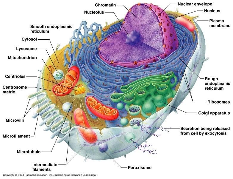
Cell: Structure and Functions (With Diagram) - Biology Discussion Eukaryotic Cells: 1. Eukaryotes are sophisticated cells with a well defined nucleus and cell organelles. 2. The cells are comparatively larger in size (10-100 μm). 3. Unicellular to multicellular in nature and evolved ~1 billion years ago. 4. The cell membrane is semipermeable and flexible. 5. These cells reproduce both asexually and sexually. A Labeled Diagram of the Animal Cell and its Organelles One can observe the golgi apparatus in the labeled animal cell parts diagram. The golgi apparatus is situated near the cell nucleus and besides the stacked sacs, it also contains large number of vesicles. The main function of this golgi complex is to receive the proteins synthesized in the ER and transform it into more complex proteins. How to draw an animal cell - labeled science diagram - YouTube Download a free printable outline of this video and draw along with us: you for watching. Please ... Structure of Cell: Definition, Types, Diagram, Functions - Embibe Structure of Cell: Cell is the basic functional unit that makes up all living organisms.All organisms, including ourselves, start life as a single cell called the egg. Cells are small microscopic units that perform all essential functions of life and are capable of independent existence.
Animal Cell Diagram PDF in 2022 - Pinterest Budding biologists, here's a great cheat-sheet to help you memorize and compare the structures of both animal cells and plant cells. A printable diagram of an animal cell. This PDF includes the color version, black and white version, and the labeled and unlabeled diagrams for students to complete. 6 pages total. Animal Cells: Labelled Diagram, Definitions, and Structure Animal Cells Organelles and Functions. A double layer that supports and protects the cell. Allows materials in and out. The control center of the cell. Nucleus contains majority of cell's the DNA. Popularly known as the "Powerhouse". Breaks down food to produce energy in the form of ATP. animal cell diagram with labels Animal plant cell - Teaching resources. 18 Pictures about Animal plant cell - Teaching resources : Animal Cell by Amy Conley, label the parts of animal cells - Brainly.in and also Questions And Answers On Labeled/Unlebled Diagrams Of A Human Cell. A Labelled Diagram Of Neuron with Detailed Explanations Diagram Of Neuron. A neuron is a specialized cell, primarily involved in transmitting information through electrical and chemical signals. They are found in the brain, spinal cord and the peripheral nerves. A neuron is also known as the nerve cell. The structure of a neuron varies with their shape and size and it mainly depends upon their ...
The Cell
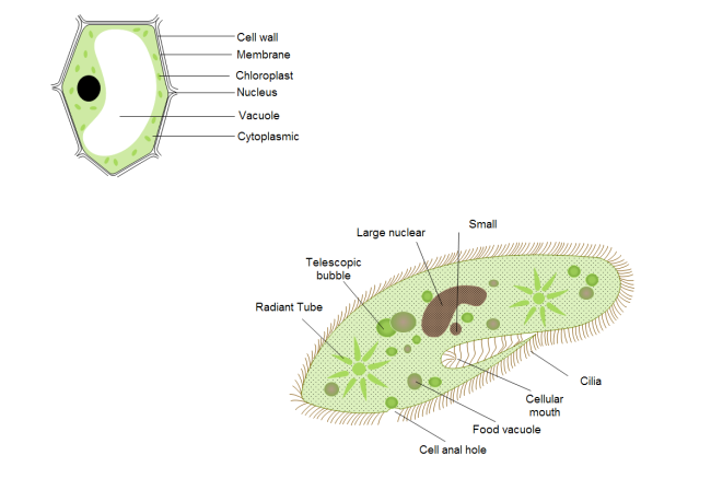
Labeling a Cell Diagram | Quizlet Cell Wall This gives shape and support to the plant cell. It surrounds the cell and protects the other parts of the cell. Chloroplasts This is where the plant cell's chlorophyll is stored. This is what the plant uses to make its own food (photosynthesis). This is also what makes plant cells have a green-like color. Plant cells Are circular in shape

Copy of "Plant cell"
03 Label the Cell Diagram | Quizlet Start studying 03 Label the Cell. Learn vocabulary, terms, and more with flashcards, games, and other study tools.
A Typical Cell, Labeled Stock Vector Illustration 112395593 : Shutterstock
Diagram of a cell membrane with labels - NIST Essential Biological FunctionsImmune response, Cell metabolism, Neurotransmission, Photosynthesis, Cell adherence, Cell growth and differentiationPotential Commercial ApplicationsDrug response monitoring, Chemical manufacturing, Biosensing, Energy conversion, Tissue engineering ... Diagram of a cell membrane with labels. Appears In. Biology in ...

The Prokaryotic Cell (Bacteria) - PurposeGames
Label Cell Parts | Plant & Animal Cell Activity | StoryboardThat Create a cell diagram with each part of plant and animal cells labeled. Include descriptions of what each organelle does. Click "Start Assignment". Find diagrams of a plant and an animal cell in the Science tab. Using arrows and Textables, label each part of the cell and describe its function.
Cell Diagram | Free Cell Diagram Templates
A Labeled Diagram of the Plant Cell and Functions of its Organelles A Labeled Diagram of the Plant Cell and Functions of its Organelles We are aware that all life stems from a single cell, and that the cell is the most basic unit of all living organisms. The cell being the smallest unit of life, is akin to a tiny room which houses several organs. Here, let's study the plant cell in detail...
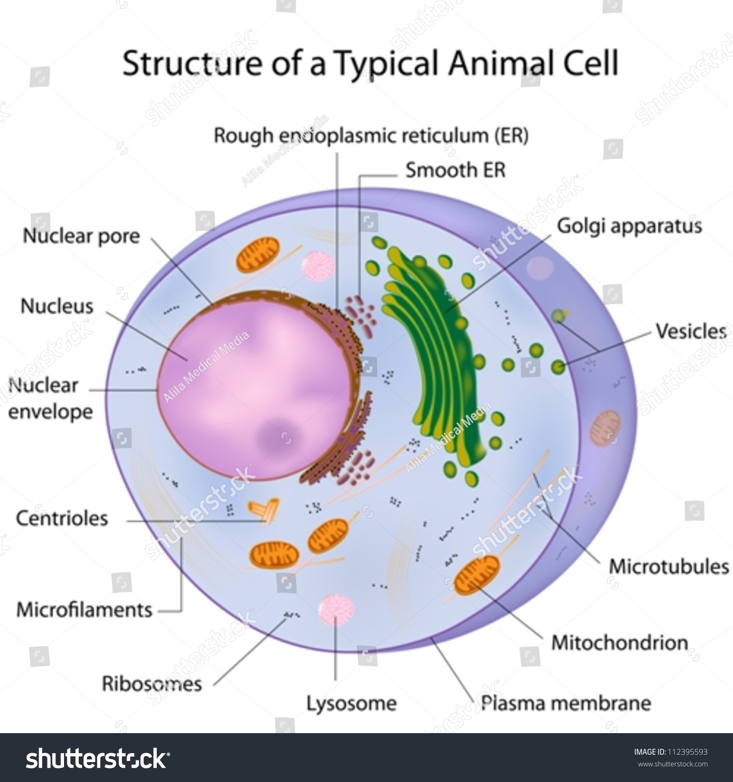
Typical Cell Labeled Stock Vector 112395593 - Shutterstock
Cells Diagram | Science Illustration Solutions - Edrawsoft Cells Diagram Symbols Edraw software offers you lots of symbols used in cells diagram like cell structure, paramecium, squamous cell, cell division, bacteria, cell membrane, eggs, sperm, zygote, an animal cell, SARS, tobacco mosaic, adenovirus, coliphage, herpesvirus, AIDS, pollen, plant cell model, onion tissue, etc. Cells Diagram Examples

HAIR REMOVAL EXPERT: plant cell diagram with labels for kids
Bacteria in Microbiology - shapes, structure and diagram Bacterial spores. Bacterial endospores layers. Bacteria cells are the smallest living cells that are known; even though viruses are smaller than bacteria, viruses are not living cells. There are different types of bacteria with various sizes, shapes, and structures. The bacteria shapes, structure, and labeled diagrams are discussed below.
Print Chapter 2 cells flashcards | Easy Notecards
Cell Organelles- Definition, Structure, Functions, Diagram In a plant cell, the cell wall is made up of cellulose, hemicellulose, and proteins while in a fungal cell, it is composed of chitin. A cell wall is multilayered with a middle lamina, a primary cell wall, and a secondary cell wall. The middle lamina contains polysaccharides that provide adhesion and allow binding of the cells to one another.
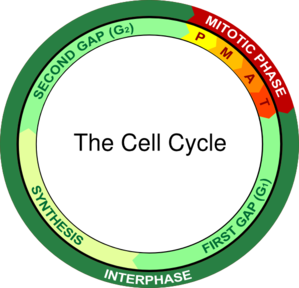
Cell Division
How to draw a nerve cell - labeled science diagrams - YouTube Download a free printable outline of this video and draw along with us: you for watching. Please su...
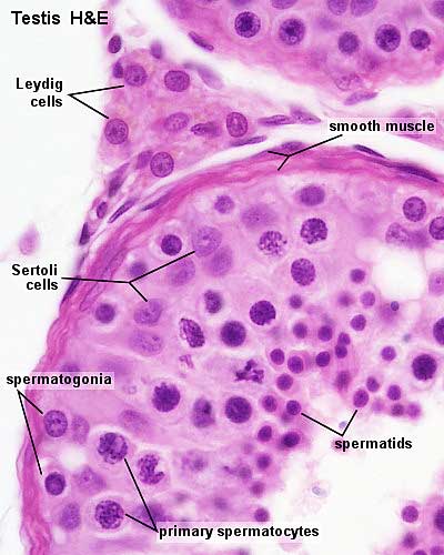
ANAT2341 Lab 1 - Spermatogenesis - Embryology
Human Cell Diagram, Parts, Pictures, Structure and Functions One of the few cells in the human body that lacks almost all organelles are the red blood cells. The main organelles are as follows : cell membrane endoplasmic reticulum Golgi apparatus lysosomes mitochondria nucleus perioxisomes microfilaments and microtubules Diagram of the human cell illustrating the different parts of the cell. Cell Membrane
Talk:ANAT2511 Introduction to Histology - Embryology
Animal Cell Diagram with Label and Explanation: Cell Structure, Functions Animal cell is a typical Eukaryotic cell enclosed by a plasma membrane containing nucleus and organelles which lack cell walls, unlike all other Eukaryotic cells. The typical cell ranges in size between 1-100 micrometers. The lack of cell walls enabled the animal cells to develop a greater diversity of cell types.
.jpg)
Chuck Whicker, Connor Wagner, Katie Wright --- Animal Cell Diagram | Loveland AP Biology
Learn the parts of a cell with diagrams and cell quizzes - Kenhub For this exercise we'll start with an image of a cell diagram ready labeled. Study this and make sure that you're clear about which structure is found where. Cell diagram unlabeled It's time to label the cell yourself! As you fill in the cell structure worksheet, remember the functions of each part of the cell that you learned in the video.
Biology 101: Cells | Owlcation
diagram of a cell labeled diagram of a cell labeled Paramecium under 400x magnification. Explain the nucleus of a cell with a neat labeled diagram. Volvox cells protist colony flagella practical lab daughter quia parent amoeba feldman 1b bio vocabulary diagram of a cell labeled
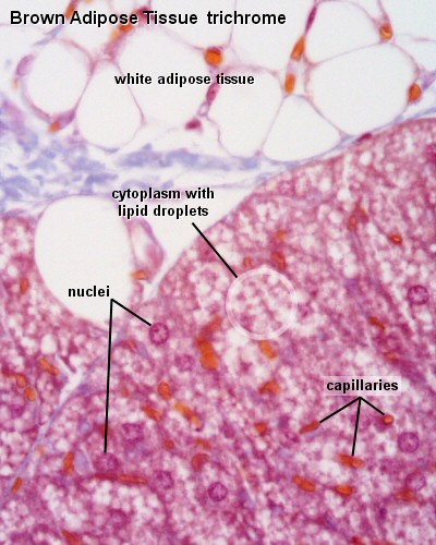
ANAT2241 Connective Tissue Components - Embryology
Ch 3 Review Guide Answer Key

Basic Plant Cell Diagram With Labels
Post a Comment for "42 diagram of a cell with labels"